...
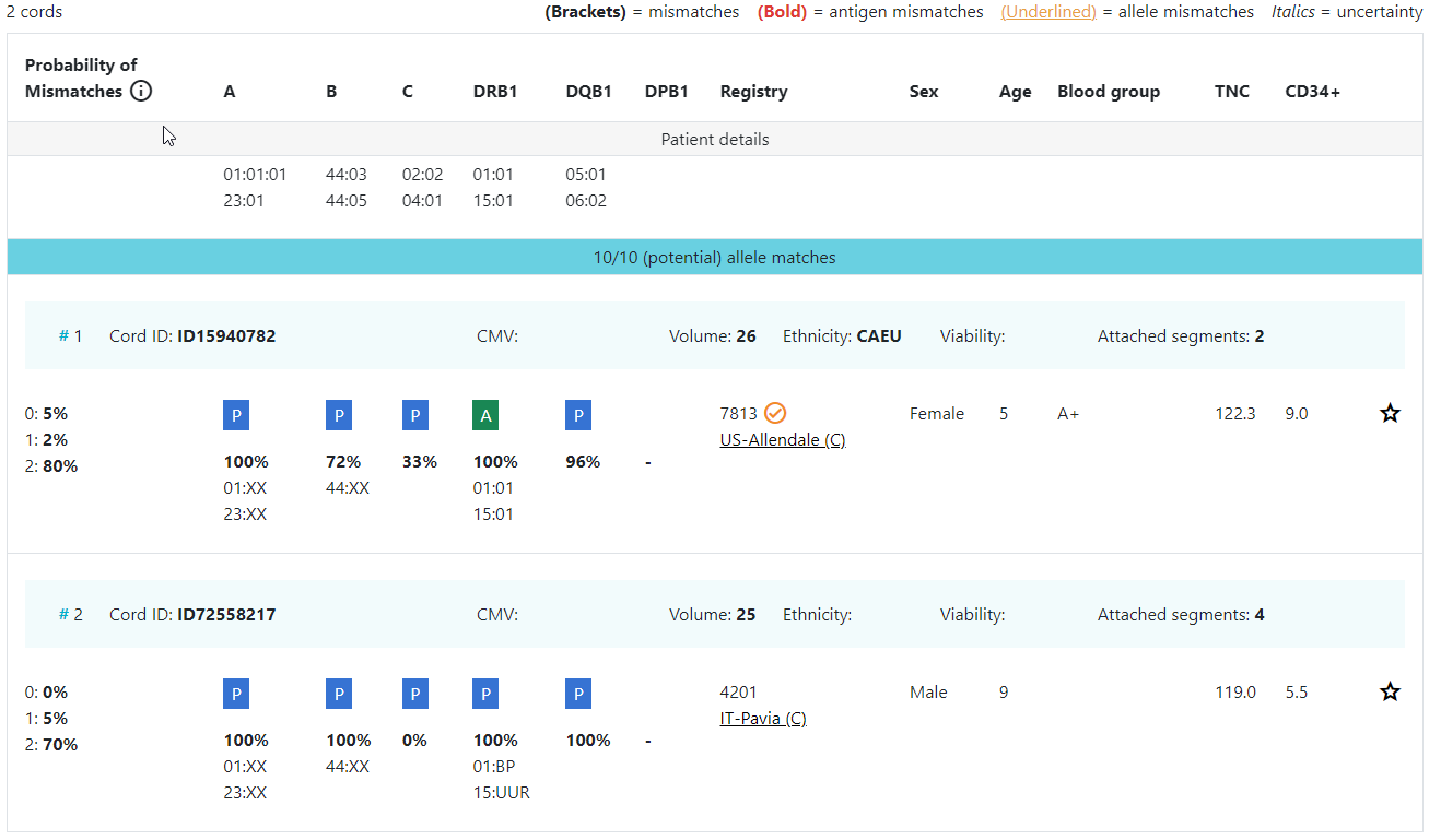
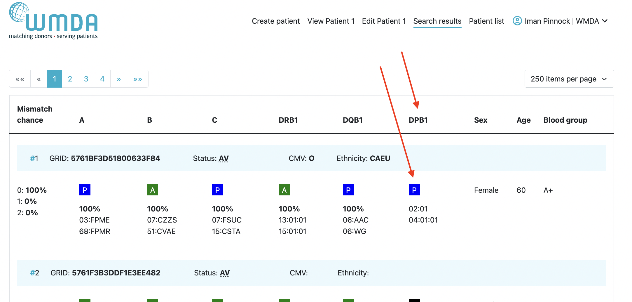
An overview of the match results table of donor search results and cord search results are shown in figure 16 and 17.
Donor search
By default the donor match results is as follows:0 mismatch search → "Standard sorting"
- HLA match grade (e.g. first 8/8 then 7/8, 6/8)
- Match probability in 10% intervals (descending)
- Donor age (ascending)
For 1 mismatch search → and 2 mismatch searches, the sorting method can be changed to "Sum of probabilities sorting"
- Sum of match probability for 0 mismatch and 1 mismatch, in 10% intervals (descending)
- Donor age (ascending)
2 mismatch search → "Sum of probabilities sorting"
...
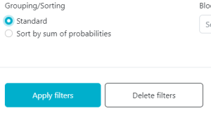
". You can do this by clicking on the "all filters" button and selecting "Sort by sum of probabilities" under "Grouping/Sorting".

CBU search
...
By default the cord match results are sorted as follows:
0 mismatch search → "Standard sorting"
- HLA match grade (e.g. first 8/8 then 7/8, 6/8)
- Match probability in 10% intervals (descending)
- Number of total nucleated cells (TNC, descending)
1 mismatch search → "Sum of probabilities sorting"
- Sum of match probability for 0 mismatch and 1 mismatch, in 10% intervals (descending)
- Number of total nucleated cells (TNC, descending)
2 mismatch search → "Sum of probabilities sorting"
- )
- Match probability Sum of match probability for 0, 1 and 2 mismatch, in 10% intervals (descending)
- Number of total nucleated cells (TNC, descending)
If the search is a mismatch search but you would still like to have the search results sorted the "standard" way, You For 1 and 2 mismatch searches, the sorting method can be changed to "Sum of probabilities sorting". You can do this by clicking on the "all filters" button and selecting "StandardSort by sum of probabilities" under "Grouping/Sorting".
Overview of donor search match results
...
| Imagefloat | ||
|---|---|---|
| ||
|
Explanation of colours, abbreviations, percentages and codes
Abbreviation / column | Description |
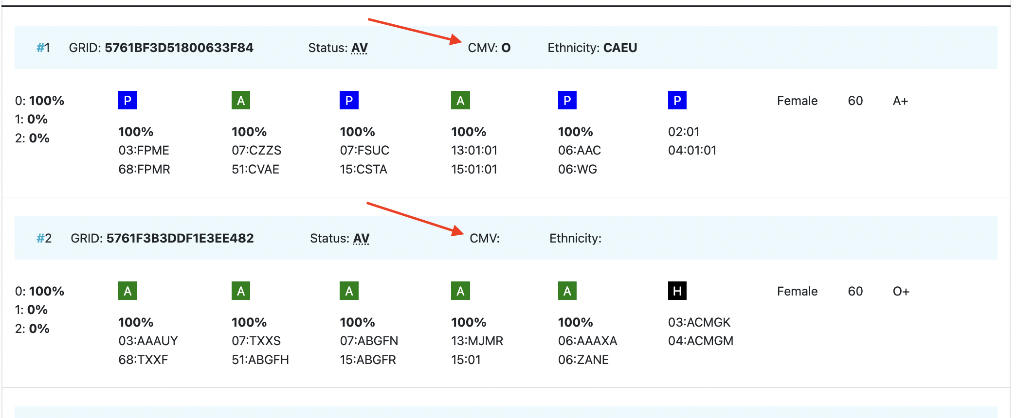
| HLA patient | In between two grey bars, in a white space, you can find the HLA of your patient. This sticky header will move with you when you are looking at results more below. |
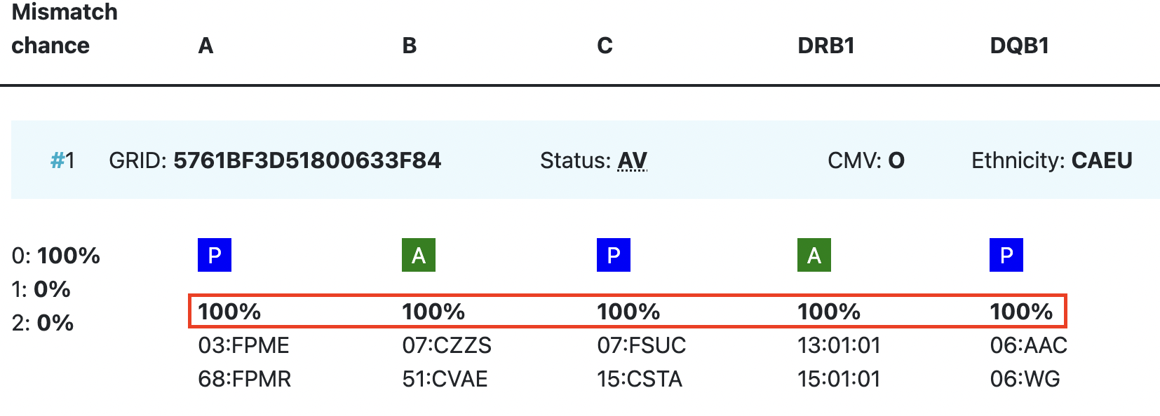
Probability of mismatches 0, 1, 2 | Probability of a mismatch at 0 loci, 1 locus, and 2 loci. The percentages are based on the match type you have been chosen (out of 6 then 3 loci are considered; out of 10 then 5 loci are considered).
The five squares above the probability percentages are representing, respectively, locus A, B, C, DRB1, and DQB1. They are showing in letter/colour codes if a certain locus of a donor/cord is likely to match with your patient or not.
|
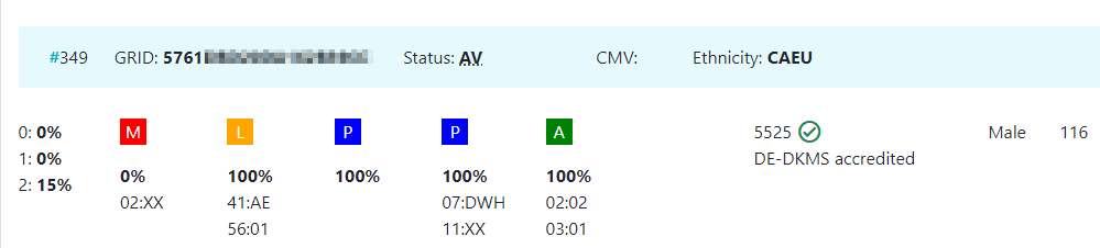
| DPB1 TCE3 grading model | The sixth square indicates the DPB1 match grade. DPB1 TCE3 evaluation is performed and displayed for potential donors under the following conditions:
The results of the DPB1 TCE3 grading is shown below the donor’s DPB1 values by using the following symbols above the DPB1 alleles of the donor:
The explanation of the symbols is also provided when hovering the symbols.
|
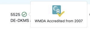
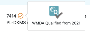
Registry Reg Abbr | This column shows you the ION code of the registry or cord blood bank where the donor or cord is registered followed by the abbreviated name.
This column may also shows you an icon that indicates that the registry is either WMDA accredited or WMDA qualified. If no icon is present next to the ION code, then the registry is not WMDA qualified or accredited.
When you hover over the icon, the icon indicates whether the registry has been WMDA qualified or whether the registry has been fully accredited. Also the validity period of the qualification/accreditation is visible. The orange ticks have verification WMDA Qualified
|
Age | Current age of donor (in case of donor search)/ Time since cryopreservation of cord blood unit (in case of cord search) |
Gender | Sex: M = male, F = female |
Blood group | Blood group, e.g. A+ = blood group A, rhesus positive, B- = blood group B, rhesus negative |
CMV | CMV status Possible values: As seen in the second entry, a value for the CMV status may not always be available.
NOTE: A tooltip for information on the values can be used to aid in understanding their meaning. |
| Probability of match per locus | Within the donor/ cord details, the probability of a match per locus is displayed:
This also correspondents with the letter/colour code from the five squares in the column probability of mismatches. These probabilities are only calculated for the 5 loci A, B, C, DRB1, and DQB1. For more information on the differences between Hap-E and Optimas please see the chapter below called "Differences between Hap-E and Optimas regarding locus specific match probabilities" |
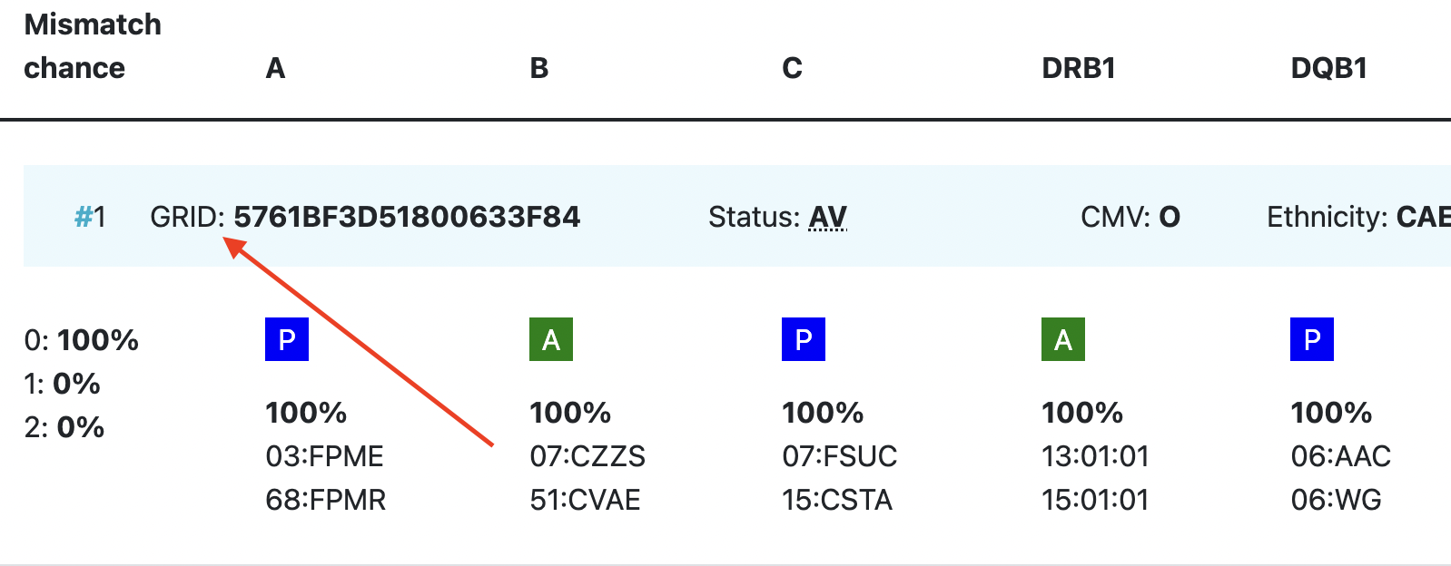
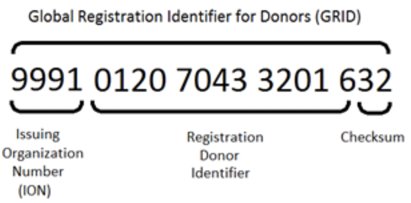
| GRID | GRID, Global Registration Identifier for Donors, is an ID for donors (not for CBUs) that is globally unique.
The number is split into 3 sections;
Once a GRID is assigned it cannot and will not be reassigned - it is completely unique.
For more information on the GRID number please refer to Global Registration Identifier for Donors - WMDA. |
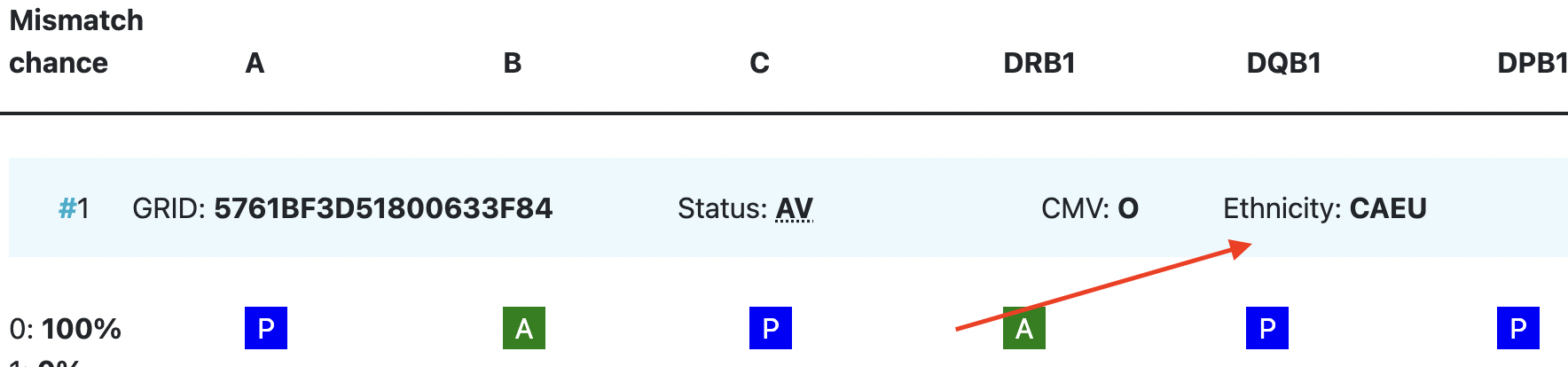
Ethnicity | Ethnic group: The system uses the same ethnic groups as defined for the EMDIS system:
The ethnic groups are as follows;
|
| Status | Status of a donor or CBU. For donors, the status can be available (AV), reserved for a patient (RS), temporarily unavailable (TU) |


...